Update: I've added details of a tentative identification feature in the basal maculation of the hindwing - at the bottom of this page:
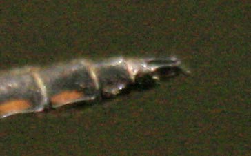
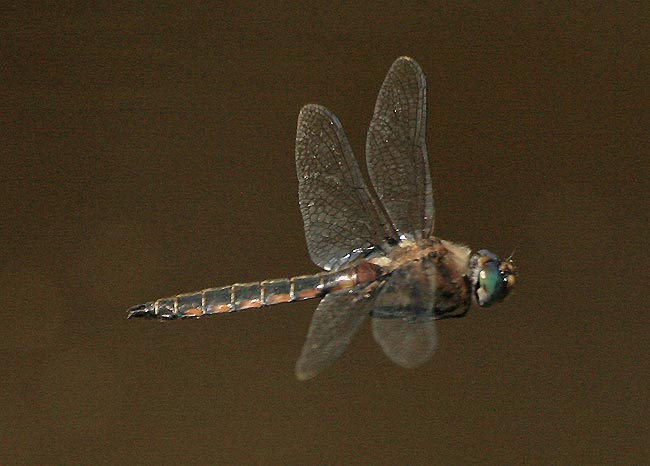
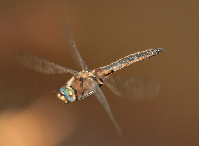
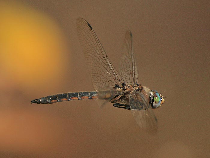
These Robust Baskettails Epitheca spinosa were photographed at Ratcliff Lake, Davy Crockett National Forest, Houston county, Texas on March 26 2008:
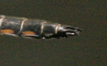
1)

2)
3)
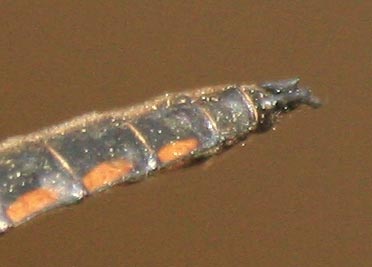
4)
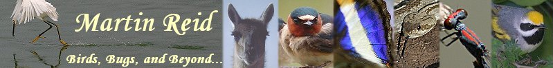
Tentative identification feature: while studying my photos and those of John Abbot and the few available on the Internet, I noticed what seems to be a consistent difference in the basal maculation on the hindwing for spinosa compared to cynsura, costalis, and petechialis:
Here's a crop of the wing of a costalis/petechialis from Fort Worth, Texas - essentially cynosura is the same:
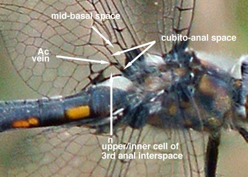
- note the following points:-
1) there is some maculation at the base of the mid-basal space - the amount is variable, but there seems to always be some present and its outer edge is at an obvious angle to the body axis.
2) the inner part of the cubito-anal space (the part inwards from Ac vein) has a variable dark inner part but always is clear distally such that the Ac vein is distinct.
3) the 3rd anal interspace of male Epitheca consists of a triangular section that is split into a larger upper cell and smaller triangular lower part (1 or 2 narrow triangular cells); the large upper cell of this space (equates to paranal cell n) is clear, and the small lower part has a variable amount of maculation (more often clear on costalis and petechialis?).
here is a profile shot to better-illustrate the above points - on this individual the dark at the base of the inner part of the cubito-anal space is smaller than on the first example such that vein Ac is clearly isolated:
Now look at spinosa:
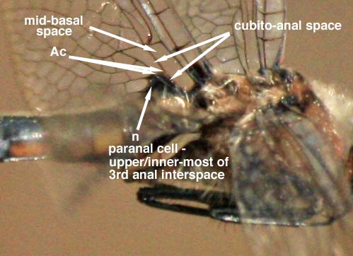
- note the following points:-
1) there is less maculation at the base of the mid-basal space - often there is no maculation at all, but when present it is very restricted and its outer edge is roughly parallel to the body axis.
2) the inner part of the cubito-anal space (the part inwards from Ac vein) is completely dark such that the Ac vein is indistinct.
3) the large upper cell of the 3rd anal interspace (equates to paranal cell n) is dark, and the small lower part has a variable amount of maculation at the upper, blunt end (can be all-dark, or with a small clear lower point).
4) the dark large upper cell of the 3rd anal interspace merges with the dark inner part of the cubito-anal space to form a contiguous triangular dark patch immediately below the mid-basal space.
- in combination this creates a distinct macular pattern to the base of the hindwing. My sample size is rather small, but thus far all examined pics seem to be consistent with the above analysis; here are some samples on the internet:
spinosa:
A male by Giff Beaton.
Another male by Giff Beaton.
A female by R. Stephen Krotzer - this is the only definite female image I could locate.
A male on Odonata Central, presumably by John Abbott.
Another male on Odonata Central, presumably by John Abbott.
others:
A petechialis from West Texas.
A selection of cynosura?/costalis?/petechialis? from Fort Worth.
A cynosura from Odonata Central.
This cynosura from Odonata Central seems to have a bit of extra maculation - more than any costalis/petechialis/spinosa?; even so, note the clear upper cell of the 3rd anal interspace. On cynosura with even more maculation, the above-described pattern may disappear beneath the extra maculation - but such individuals are not confusable with spinosa.
I'd be most keen to get feedback from those with extensive photos/specimens to draw upon - thanks.